
按品牌选择
 |
|
|||||||||||||||||||||||||||||||||
| [发表评论] [本类其他产品] [本类其他供应商] [收藏] | ||||||||||||||||||||||||||||||||||
| 销售商: 上海语纯生物科技有限公司 | 查看该公司所有产品 >> |
TUNEL细胞凋亡检测试剂盒
Kit Components:
Introduction:
DNA fragmentation represents a characteristic of late stage apoptosis. DNA fragmentation in apoptotic cells can be detected by terminal deoxynucleotidyl transferase (TdT)-mediated dUTP nick end labeling (TUNEL). The TUNEL assay relies on the presence of nicks in the DNA which can be identified by TdT, an enzyme that catalyzes the addition of dUTPs that are secondarily labeled with a marker. All the existing TUNEL assays contain the highly toxic sodium cacodylate which might induces apoptosis and also decrease DNA production and DNA strands. Our Cell Meter™ TUNEL Apoptosis Assay Kit uses proprietary buffer system free of sodium cacodylate. The kit is based on incorporation of a red fluorescence dye modified deoxyuridine 5'-triphosphates at the 3' OH ends of the DNA fragments that form during apoptosis. The assay is optimized for the direct detection of apoptosis in either detached or attached cells without using antibody.
The kit provides all the essential components with an optimized assay protocol. It is suitable for fluorescence microplate reader, fluorescence microscope, or flow cytometer. Its signal can be detected with a TRITC filter set or at Ex/Em = 550 nm/590-650 nm.
Assay Protocol:
2. Fixation and Permeabilization
2.1 Remove cell media.
2.2 Add 100μL/well/96-well plate of 4% formaldehyde fixative buffer (not supplied) to each well.
Note: For non-adherent cells, add desired amount (such as 2 x 10⁶ cells/mL) of 4% formaldehyde fixative buffer.
2.3 Incubate plates for 20 to 30 minutes at room temperature.
2.4 Remove fixative.
Optional: add 100μL/well/96-well plate of the permeabilization reagent (0.2% Triton X-100 in PBS, not supplied) after the fixation if needed, and incubate the plate for 10 minutes at room temperature.
2.5 Wash the cells with PBS 2-3 times.
Optional: You may also prepare a positive control for TUNEL reaction using DNAase I by digesting cells with DNAase I for 30 min at room temperature before proceed to TUNEL reaction (Step 3)
3. TUNEL reaction
3.1 Prepare reaction mixture just before use based on the number of samples to be assayed:
Note: Each cell line should be evaluated on an individual basis to determine the optimal cell density.
3.2 Add 50μL of the reaction mixture (from Step 3.1) to each well or tube and incubate at 37℃ for 60 minutes.
3.3 Remove the reaction mixture, and wash the cells 3-5 times with 200μL /well of PBS.
4. Monitor the fluorescence intensity by fluorescence microscope, flow cytometer, or fluorescence microplate reader at Ex/Em = 550/590-650 nm.
5. Optional: Stain the nucleus with 1× Hoechst (Component C, Ex/Em = 350/460 nm) for image analysis.
Data Analysis:
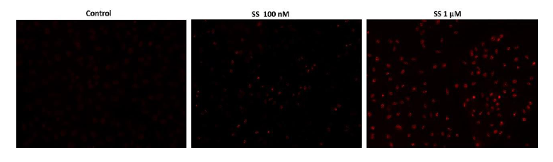
Figure 1. Fluorescence images of TUNEL reaction in HeLa cells with the treatment of 100 nM or 1μM staurosporin (SS) for 4 hours as compare to untreated control. Cells were incubated with reaction mixture for 1 hour at 37ºC. The red fluorescence signal was analyzed using fluorescence microscope with a TRITC filter set. Fluorescently labeled DNA strand breaks shows intense fluorescent staining in SS treated cells.
Kit Components:
| Components | Amount |
| Component A: 100×TunnelyteTM Red | 1 vial (25 uL) |
| Component B: Reaction Buffer | 1 bottle (5 mL) |
| Component C: 1000×Hoechst | 1 vial (50 uL) |
DNA fragmentation represents a characteristic of late stage apoptosis. DNA fragmentation in apoptotic cells can be detected by terminal deoxynucleotidyl transferase (TdT)-mediated dUTP nick end labeling (TUNEL). The TUNEL assay relies on the presence of nicks in the DNA which can be identified by TdT, an enzyme that catalyzes the addition of dUTPs that are secondarily labeled with a marker. All the existing TUNEL assays contain the highly toxic sodium cacodylate which might induces apoptosis and also decrease DNA production and DNA strands. Our Cell Meter™ TUNEL Apoptosis Assay Kit uses proprietary buffer system free of sodium cacodylate. The kit is based on incorporation of a red fluorescence dye modified deoxyuridine 5'-triphosphates at the 3' OH ends of the DNA fragments that form during apoptosis. The assay is optimized for the direct detection of apoptosis in either detached or attached cells without using antibody.
The kit provides all the essential components with an optimized assay protocol. It is suitable for fluorescence microplate reader, fluorescence microscope, or flow cytometer. Its signal can be detected with a TRITC filter set or at Ex/Em = 550 nm/590-650 nm.
Assay Protocol:
- Culture cells to an optimal density for apoptosis induction according to your specific protocol. We recommend about 30,000 to 50,000 cells/well for adherent cells grown in a 96-well microplate culture, or about 1 to 2 x 10⁶ cells/mL for non-adherent cells. At the same time, culture a non-induced negative control cell population at the same density as the induced population for every labeling condition.
2. Fixation and Permeabilization
2.1 Remove cell media.
2.2 Add 100μL/well/96-well plate of 4% formaldehyde fixative buffer (not supplied) to each well.
Note: For non-adherent cells, add desired amount (such as 2 x 10⁶ cells/mL) of 4% formaldehyde fixative buffer.
2.3 Incubate plates for 20 to 30 minutes at room temperature.
2.4 Remove fixative.
Optional: add 100μL/well/96-well plate of the permeabilization reagent (0.2% Triton X-100 in PBS, not supplied) after the fixation if needed, and incubate the plate for 10 minutes at room temperature.
2.5 Wash the cells with PBS 2-3 times.
Optional: You may also prepare a positive control for TUNEL reaction using DNAase I by digesting cells with DNAase I for 30 min at room temperature before proceed to TUNEL reaction (Step 3)
3. TUNEL reaction
3.1 Prepare reaction mixture just before use based on the number of samples to be assayed:
| Reaction Components | Volume Per Well |
| 100×TunnelyteTM Red(Component A) | 0.5μL |
| Reaction Buffer(Component B) | 50μL |
| Total volume | 50.5μL |
3.2 Add 50μL of the reaction mixture (from Step 3.1) to each well or tube and incubate at 37℃ for 60 minutes.
3.3 Remove the reaction mixture, and wash the cells 3-5 times with 200μL /well of PBS.
4. Monitor the fluorescence intensity by fluorescence microscope, flow cytometer, or fluorescence microplate reader at Ex/Em = 550/590-650 nm.
5. Optional: Stain the nucleus with 1× Hoechst (Component C, Ex/Em = 350/460 nm) for image analysis.
Data Analysis:
Figure 1. Fluorescence images of TUNEL reaction in HeLa cells with the treatment of 100 nM or 1μM staurosporin (SS) for 4 hours as compare to untreated control. Cells were incubated with reaction mixture for 1 hour at 37ºC. The red fluorescence signal was analyzed using fluorescence microscope with a TRITC filter set. Fluorescently labeled DNA strand breaks shows intense fluorescent staining in SS treated cells.
手机版:TUNEL细胞凋亡检测试剂盒


Copyright(C) 1998-2025 生物器材网 电话:021-64166852;13621656896 E-mail:info@bio-equip.com