
 |
|
|||||||||||||||||||||||||||||||||
| [发表评论] [本类其他产品] [本类其他供应商] [收藏] | ||||||||||||||||||||||||||||||||||
| 销售商: 上海雅吉生物科技有限公司 | 查看该公司所有产品 >> |
英文名称 AKT1
中文名称 蛋白激酶B重组兔单克隆抗体
别 名 AKT 1; AKT; AKT1; AKT-1; AKT1_HUMAN; C AKT; cAKT; MGC9965; MGC99656; Oncogene AKT1; PKB; PKB alpha; PKB-ALPHA; PRKBA; Protein Kinase B Alpha; Protein kinase B; Proto-oncogene c-Akt; RAC Alpha; RAC alpha serine/threonine protein kinase; RAC; RAC PK Alpha; Rac protein kinase alpha; RAC Serine/Threonine Protein Kinase; RAC-alpha serine/threonine-protein kinase; RAC-PK-alpha; v akt murine thymoma viral oncogene homolog 1; vAKT Murine Thymoma Viral Oncogene Homolog 1.
研究领域 肿瘤 细胞生物 神经生物学 信号转导 细胞凋亡 激酶和磷酸酶
抗体来源 Rabbit
克隆类型 Monoclonal
克 隆 号 11F2
交叉反应 Human, Mouse, Rat,
产品应用 WB=1:500-2000 IP=1:10-50 IHC-P=1:50-200 IHC-F=1:50-200 ICC=1:50-100 IF=1:50-100 (石蜡切片需做抗原修复)
not yet tested in other applications.
optimal dilutions/concentrations should be determined by the end user.
分 子 量 56kDa
细胞定位 细胞核 细胞浆 细胞膜
性 状 Liquid
浓 度 1mg/ml
免 疫 原 Recombinant human AKT1 protein:
亚 型 IgG
纯化方法 affinity purified by Protein A
储 存 液 0.01M TBS(pH7.4) with 1% BSA, 0.03% Proclin300 and 50% Glycerol.
保存条件 Shipped at 4℃. Store at -20 °C for one year. Avoid repeated freeze/thaw cycles.
PubMed PubMed
产品介绍 The serine-threonine protein kinase encoded by the AKT1 gene is catalytically inactive in serum-starved primary and immortalized fibroblasts. AKT1 and the related AKT2 are activated by platelet-derived growth factor. The activation is rapid and specific, and it is abrogated by mutations in the pleckstrin homology domain of AKT1. It was shown that the activation occurs through phosphatidylinositol 3-kinase. In the developing nervous system AKT is a critical mediator of growth factor-induced neuronal survival. Survival factors can suppress apoptosis in a transcription-independent manner by activating the serine/threonine kinase AKT1, which then phosphorylates and inactivates components of the apoptotic machinery. Mutations in this gene have been associated with the Proteus syndrome. Multiple alternatively spliced transcript variants have been found for this gene. [provided by Ref
| 产品图片 | 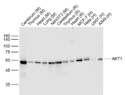 Sample: Lane 1: Cerebrum (Mouse) Lysate at 40 ug Lane 2: Thymus (Mouse) Lysate at 40 ug Lane 3: Heart (Mouse) Lysate at 40 ug Lane 4: Lung (Mouse) Lysate at 40 ug Lane 5: NIH/3T3 (Mouse) Cell Lysate at 30 ug Lane 6: Cerebellum (Rat) Lysate at 40 ug Lane 7: Thymus (Rat) Lysate at 40 ug Lane 8: Heart (Rat) Lysate at 40 ug Lane 9: MCF-7 (Human) Cell Lysate at 30 ug Lane 10: Hela (Human) Cell Lysate at 30 ug Lane 11: U937 (Human) Cell Lysate at 30 ug Lane 12: A549 (Human) Cell Lysate at 30 ug Primary: Anti- AKT1 (bsm-52010R) at 1/1000 dilution Secondary: IRDye800CW Goat Anti-Rabbit IgG at 1/20000 dilution Predicted band size: 59 kD Observed band size: 59 kD  Sample: Cerebrum (Mouse) Lysate at 40 ug Lung (Mouse) Lysate at 40 ug Heart (Mouse) Lysate at 40 ug Hela(Human) Cell Lysate at 30 ug NIH/3T3(Mouse) Cell Lysate at 30 ug A549(Human) Cell Lysate at 30 ug HepG2(Human) Cell Lysate at 30 ug Primary: Anti- AKT1 (bsm-52010R) at 1/1000 dilution Secondary: IRDye800CW Goat Anti-Rabbit IgG at 1/20000 dilution Predicted band size: 56 kD Observed band size: 56 kD  Paraformaldehyde-fixed, paraffin embedded (rat brain); Antigen retrieval by boiling in sodium citrate buffer (pH6.0) for 15min; Block endogenous peroxidase by 3% hydrogen peroxide for 20 minutes; Blocking buffer (normal goat serum) at 37°C for 30min; Antibody incubation with (AKT1) Monoclonal Antibody, Unconjugated (bsm-52010R) at 1:200 overnight at 4°C, followed by operating according to SP Kit(Rabbit) (sp-0023) instructionsand DAB staining.  Paraformaldehyde-fixed, paraffin embedded (mouse brain); Antigen retrieval by boiling in sodium citrate buffer (pH6.0) for 15min; Block endogenous peroxidase by 3% hydrogen peroxide for 20 minutes; Blocking buffer (normal goat serum) at 37°C for 30min; Antibody incubation with (AKT1) Monoclonal Antibody, Unconjugated (bsm-52010R) at 1:200 overnight at 4°C, followed by operating according to SP Kit(Rabbit) (sp-0023) instructionsand DAB staining. 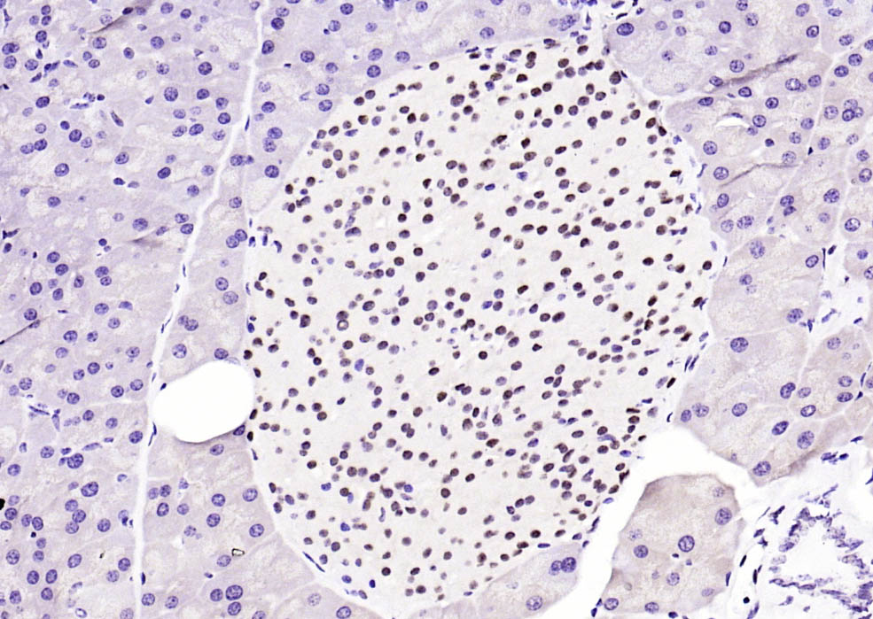 Paraformaldehyde-fixed, paraffin embedded (rat pancreas); Antigen retrieval by boiling in sodium citrate buffer (pH6.0) for 15min; Block endogenous peroxidase by 3% hydrogen peroxide for 20 minutes; Blocking buffer (normal goat serum) at 37°C for 30min; Antibody incubation with (AKT1) Monoclonal Antibody, Unconjugated (bsm-52010R) at 1:200 overnight at 4°C, followed by operating according to SP Kit(Rabbit) (sp-0023) instructionsand DAB staining.  Paraformaldehyde-fixed, paraffin embedded (rat heart); Antigen retrieval by boiling in sodium citrate buffer (pH6.0) for 15min; Block endogenous peroxidase by 3% hydrogen peroxide for 20 minutes; Blocking buffer (normal goat serum) at 37°C for 30min; Antibody incubation with (AKT1) Monoclonal Antibody, Unconjugated (bsm-52010R) at 1:200 overnight at 4°C, followed by operating according to SP Kit(Rabbit) (sp-0023) instructionsand DAB staining.  Paraformaldehyde-fixed, paraffin embedded (rat lung); Antigen retrieval by boiling in sodium citrate buffer (pH6.0) for 15min; Block endogenous peroxidase by 3% hydrogen peroxide for 20 minutes; Blocking buffer (normal goat serum) at 37°C for 30min; Antibody incubation with (AKT1) Monoclonal Antibody, Unconjugated (bsm-52010R) at 1:200 overnight at 4°C, followed by operating according to SP Kit(Rabbit) (sp-0023) instructionsand DAB staining. 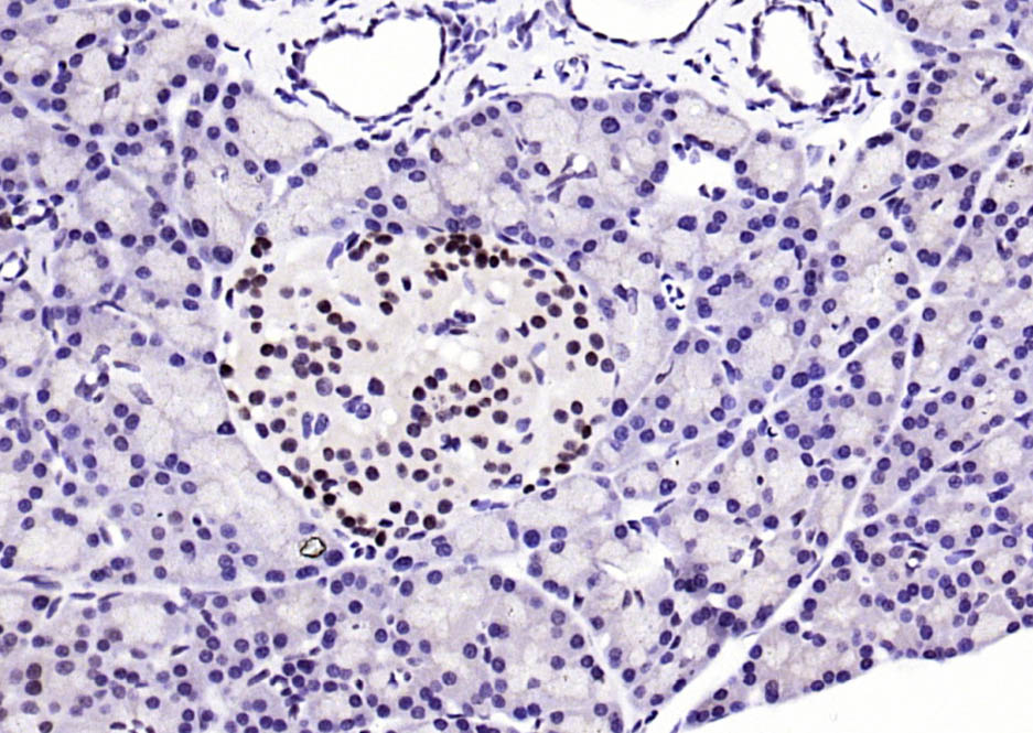 Paraformaldehyde-fixed, paraffin embedded (mouse pancreas); Antigen retrieval by boiling in sodium citrate buffer (pH6.0) for 15min; Block endogenous peroxidase by 3% hydrogen peroxide for 20 minutes; Blocking buffer (normal goat serum) at 37°C for 30min; Antibody incubation with (AKT1) Monoclonal Antibody, Unconjugated (bsm-52010R) at 1:200 overnight at 4°C, followed by operating according to SP Kit(Rabbit) (sp-0023) instructionsand DAB staining. 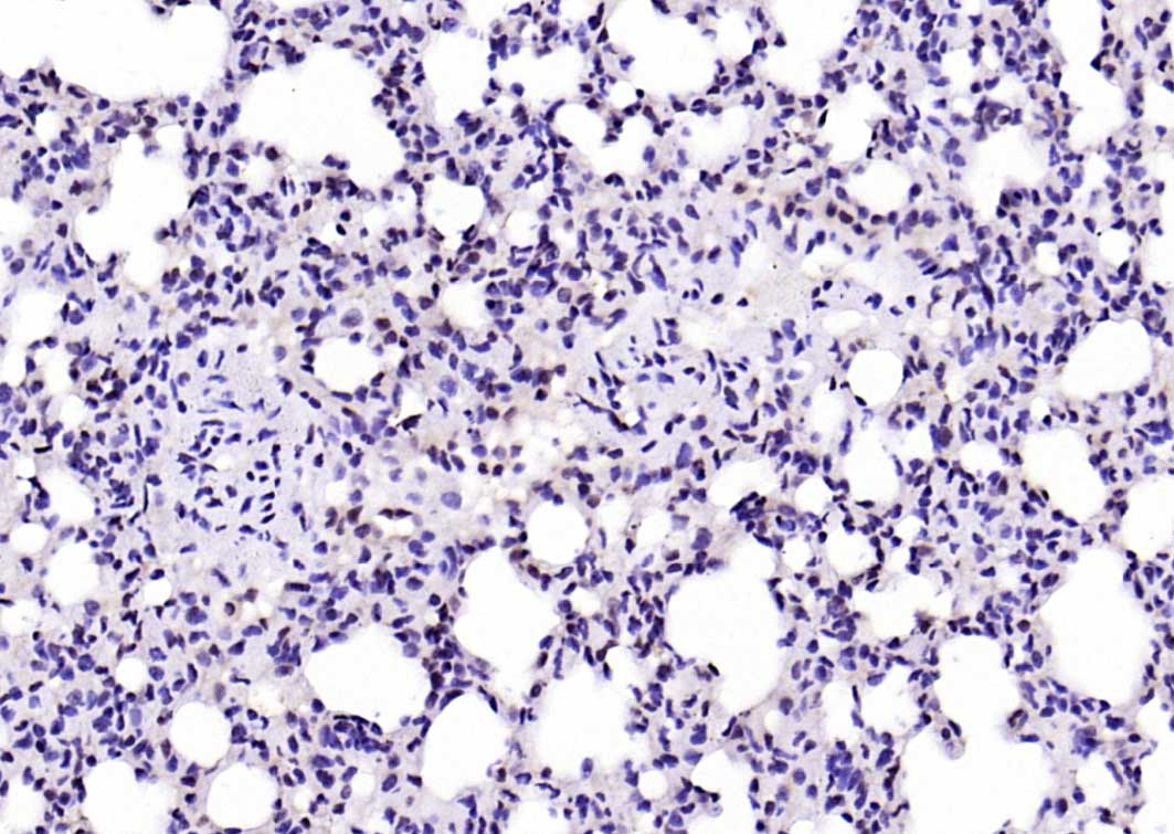 Paraformaldehyde-fixed, paraffin embedded (mouse lung); Antigen retrieval by boiling in sodium citrate buffer (pH6.0) for 15min; Block endogenous peroxidase by 3% hydrogen peroxide for 20 minutes; Blocking buffer (normal goat serum) at 37°C for 30min; Antibody incubation with (AKT1) Monoclonal Antibody, Unconjugated (bsm-52010R) at 1:200 overnight at 4°C, followed by operating according to SP Kit(Rabbit) (sp-0023) instructionsand DAB staining. |
试剂盒售后:本公司出售的试剂盒均保质保量,质量问题均可免费退换,另提供免费代测服务。
细胞售后:
1. 细胞运输丢失、瓶身破损、培养液严重漏液等,重发;
3. 细胞收到当天以及第2,3天请拍照,未告知的视为产品合格。4-10天内出现问题,请提供细胞照片和细胞出现问题的照片以及细胞相关操作的详细步骤,并跟我公司人员及时沟通判定是否重发,具体可以参照我官网或者随货说明书相关售后条款。
细胞售后:
1. 细胞运输丢失、瓶身破损、培养液严重漏液等,重发;
3. 细胞收到当天以及第2,3天请拍照,未告知的视为产品合格。4-10天内出现问题,请提供细胞照片和细胞出现问题的照片以及细胞相关操作的详细步骤,并跟我公司人员及时沟通判定是否重发,具体可以参照我官网或者随货说明书相关售后条款。
手机版:蛋白激酶B重组兔单克隆抗体


Copyright(C) 1998-2025 生物器材网 电话:021-64166852;13621656896 E-mail:info@bio-equip.com