
 |
|
|||||||||||||||||||||||||||||
| [��������] [����������Ʒ] [����������Ӧ��] [�ղ�] | ||||||||||||||||||||||||||||||
| �����̣� �Ϻ����п�ѧ��������˾ | �鿴�ù�˾���в�Ʒ >> |
�����嶨λ�������ʡ�����������ҩ��������Ƶ������ڵ���Ҫ�о��豸�������嶨λ�����ڶ��ṹ���ж����ע�䡢�̼����ƻ��������缫�Ȳ���������������ɭ�ϲ�����ģ�ͽ�������ﶯ��ģ�ͽ�������������ģ�ͽ�����ѧϰ���䣬������ϸ����ֲ����ȱѪ���о���
�����嶨λ�������ô�С���������ǰض�㣬��Bragma�㣬�������ο������涨����ά����ϵͳ,��ȷ��Ƥ����ijЩ�ṹ��λ��,ͨ���̶������嶨λ�Dz������ϵ������ض���ά������ṹ��λ��,��״��ǣ��Ա��ڷ�ֱ�ӱ�¶�¶�����ж���Ĵ̼����ƻ���ע��ҩ�������λ���о���
�����ͺŵ������嶨λ�ǣ���ֱ�۵���ʾ����λ�ǵ���ά���꣬�����������㣬�ƶ������ۺ���ʾ�ض�λ�õ��µ����꣬ͨ��ѡ�䲻ͬ���������������ڲ�ͬ��С����ʵ�顣
���������嶨λ�Dz�Ʒ�ص㣺
��������㣬���������������
�����嶨λ�DZ�����ɼ����̣�����������ȷ��Ϊ0.1mm��
�����嶨λ�Dz������ƶ���Χ�����£����ң�ǰ���������ƶ�����80mm��
��ֱ�����90��ת��������ʱ����λ�ã�
����������ǿ�������Ӳ����ۣ�����ע��װ�ü���ȣ�
���Ը�����Ҫ���Ӳ�ͬ�Ĺ̶��������ڶ��ֶ��
�����������ƣ�
· �������
· �ƶ�ƽ��
· ȫ��λ���ڵ�������������
· �����������ѡ����ֶ������������������Լ���
��������ͬ���ж��ֲ�ͬ���ͺſɹ�ѡ�����ͣ�˫���ͣ������ͣ������ͣ�����������ѯ��
�����ͺſɹ�ѡ��
· 51600���������嶨λ�ǣ�����
· 51650�ͣ��������嶨λ�ǣ��������Ǵ��̶��ˣ�
· 51603�ͣ�˫�۱������嶨λ�ǣ�����
· 51653�ͣ�˫�۱������嶨λ�ǣ��������Ǵ��̶��ˣ�
· 51600U�ͣ������ܱ������嶨λ�ǣ�����
· 51650U�ͣ������ܱ������嶨λ�ǣ������������˶��ˣ�
· 51653U�ͣ�������˫�۱������嶨λ�ǣ������������˶��ˣ�
· 51900�ͣ� ���Ա������嶨λ�ǣ�����
· 51950�����Ա������嶨λ�ǣ��������Ǵ��̶��ˣ�
· 51903������˫�۱������嶨λ�ǣ�����
· 51953������˫�۱������嶨λ�ǣ��������Ǵ��̶��ˣ�
· 51600M���綯�������嶨λ�ǣ�����
· 51601����λ�ǻ���������ͷ���̶���
�����嶨λ�ǵ���Ҫ���죺

51600���������嶨λ�ǣ�����

51900�ͣ� ���Ա������嶨λ�ǣ�����

51903������˫�۱������嶨λ�ǣ�����

SA-103�ͱ�Яʽ����λ�ǣ�

51730 �� ��ЯʽС��λ�ǣ�

51730�ͱ�Я��С��������ʾ ��λ�ǣ�

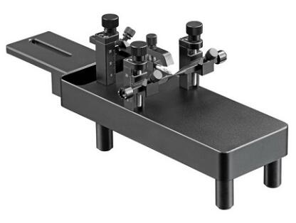
С���״��������嶨λ��������
��С�����������˲�����֬���ϣ��Լ�˽����ʺϵ��ȴ������ܹ��ι̵ļн�С��ͷ���ֱ����˲��ò������Ϊ���˶�С���ǵ����ˣ�������˵ĸ߶Ⱥ��ųݼеĸ߶Ⱦ������ɽ��е��ڣ������п̶ȣ��ʺϲ�ͬ�ĽǶȽ���ʵ�顣
����������
· �����������������嶨λ�ǵı����ã�ͨ��һ�Զ��ˡ��Ǻ��ųݼй̶�����ͷ��
· ����ͨ����β�������ƶ� 30mm
· ������100um�ľ��ȣ�ˮƽ������Դﵽ50mm���ƶ����룬���㲻ͬ���ش���Ĺ̶�
· ����������Ĭ������Ϊ18�ȶ��˺��ųݼУ�����ѡ��45�ȶ���

����С�������Ͷ��ˣ�

60° С�����


С���������嶨λ�Dz��ֲο����ף�
1. Albéri, L., Lintas, A., Kretz, R., Schwaller, B., & Villa, A. E. (2013). The calcium-binding protein parvalbumin modulates the firing 1 properties of the reticular thalamic nucleus bursting neurons. Journal of neurophysiology, 109(11), 2827-2841.
2. Sonati, T., Reimann, R. R., Falsig, J., Baral, P. K., O’Connor, T., Hornemann, S., Aguzzi, A. (2013). The toxicity of antiprion antibodies is mediated by the flexible tail of the prion protein. Nature, 501(7465), 102-106.
3. Ali, I., O’Brien, P., Kumar, G., Zheng, T., Jones, N. C., Pinault, D., O’Brien, T. J. (2013). Enduring Effects of Early Life Stress on Firing Patterns of Hippocampal and Thalamocortical Neurons in Rats: Implications for Limbic Epilepsy. PLOS ONE, 8(6), e66962.
4. Bell, L. A., Bell, K. A., & McQuiston, A. R. (2013). Synaptic Muscarinic Response Types in Hippocampal CA1 Interneurons Depend on Different Levels of Presynaptic Activity and Different Muscarinic Receptor Subtypes. Neuropharmacology.
5. Bolzoni, F., Bączyk, M., & Jankowska, E. (2013). Subcortical effects of transcranial direct current stimulation (tDCS) in the rat. The Journal of Physiology.
6. Bolzoni, F., Bączyk, M., & Jankowska, E. (2013). Subcortical effects of transcranial direct current stimulation (tDCS) in the rat. The Journal of Physiology.
7. Babaei, P., Tehrani, B. S., & Alizadeh, A. (2013). Effect of BDNF and adipose derived stem cells transplantation on cognitive deficit in Alzheimer model of rats. Journal of Behavioral and Brain Science, 3, 156-161.
8. Gilmartin, M. R., Miyawaki, H., Helmstetter, F. J., & Diba, K. (2013). Prefrontal Activity Links Nonoverlapping Events in Memory. The Journal of Neuroscience, 33(26), 10910-10914.
9. Feng, L., Sametsky, E. A., Gusev, A. G., & Uteshev, V. V. (2012). Responsiveness to nicotine of neurons of the caudal nucleus of the solitary tract correlates with the neuronal projection target. Journal of Neurophysiology, 108(7), 1884-1894.
10. Clarner, T., Diederichs, F., Berger, K., Denecke, B., Gan, L., Van der Valk, P., Kipp, M. (2012). Myelin debris regulates inflammatory responses in an experimental demyelination animal model and multiple sclerosis lesions. Glia, 60(10), 1468-1480.
11. Girardet, C., Bonnet, M. S., Jdir, R., Sadoud, M., Thirion, S., Tardivel, C., Troadec, J. D. (2011). Central inflammation and sickness-like behavior induced by the food contaminant deoxynivalenol: A PGE2-independent mechanism.Toxicological Sciences, 124(1), 179-191.
12. Hruška-Plochá��, M., Juhas, S., Juhasova, J., Galik, J., Miyanohara, A., Marsala, M., Motlik, J. (2010). A27 Expression of the human mutant huntingtin in minipig striatum induced formation of EM48+ inclusions in the neuronal nuclei, cytoplasm and processes. Journal of Neurology, Neurosurgery & Psychiatry, 81(Suppl 1), A9-A9.
13. Brooks, S., Jones, L., & Dunnett, S. B. (2010). A29 Frontostriatal pathology in the (C57BL/6J) YAC128 mouse uncovered by the operant delayed alternation task. Journal of Neurology, Neurosurgery & Psychiatry, 81(Suppl 1), A9-A10.
14. Yu, L., Metzger, S., Clemens, L. E., Ehrismann, J., Ott, T., Gu, X., Nguyen, H. P. (2010). A28 Accumulation and aggregation of human mutant huntingtin and neuron atrophy in BAC-HD transgenic rat. Journal of Neurology, Neurosurgery & Psychiatry, 81(Suppl 1), A9-A9.
15. Baxa, M., Juhas, S., Pavlok, A., Vodicka, P., Juhasova, J., Hruška-Plochá��, M., Motlik, J. (2010). A26 Transgenic miniature pig as an animal model for Huntington’s disease. Journal of Neurology, Neurosurgery & Psychiatry, 81(Suppl 1), A8-A9.
�����˽������ϸ������
����������ϵ��
TEL��021-35183767��18502129044
QQ��3007536621
Mail��yuyan0317@126.com
��ӭ������ѯ��

רҵ���ۺ�������
�ڱ������ڣ�������Ϊ���ص��������Ĺ��Ͼ����ά�����ۺ�������Ľ�Ϊ�û���Ѹ������������ȡ�κη��ã�����������Ϊ��1��


- ���ܶ��������������ڱ����С�������ʵ��⨺�ȫ������Ӧ��
- ���������ҩ�������Ҹ��ƾ�ǰ����֢���弰����֢״������
- ��ɭ�����������嶨λ��ά���ͱ���˵����
- scRNA-seq��scStereo-seq���Ͻ������Զ�����ʱ�շ���ת¼��ͼ��
- �����嶨λ���ڽ�ʾ�Ȱ�ͪ����ʱ�̡������������û����е�Ӧ��
- ���������嶨λ�����������
- ���ֵ�����2023���һ������ѧ�о��ɹ�����
- �����嶨λע��ϵͳ�ڽ�ʾ���ν���ϸ�����ڿɿ������»����е�Ӧ��